en English
Untitled English subtitles
Report a problem
Advertisement
English transcript
Gram Staining
The gram stain is one of the most useful methods in identifying bacteria.
It is an example for differential staining technique.
In this procedure, the bacteria is stained and examined under the microscope.
Gram stain permits the separation of all bacteria into two large groups, those which retain
the primary dye (gram-positive) and those that take the colour of the counter stain
(gram-negative).
Procedure:
Heat the inoculation loop with burner. Start burning the loop horizontally from its base
to vertical position till it becomes red hot. Touch an isolated colony with inoculating
loop and swirl it in the drop of water on the slide.
If it is broth culture, a loopful of culture can be placed directly on a clean microscopic
slide. Now,let the smear air dry at room temperature
Now, fix the smear using heat by waving the slide over a flame and be careful not to overheat.
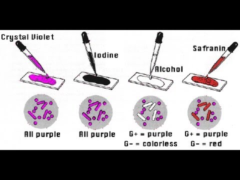
Flood the slide with crystal violet, and let stand for one minute.
Wash the slide briefly with cold water. Flood the slide with gram's iodine; let stand
for one minute; wash off with water. Decolorize until the water flows colourlessly
from the slide. Now, flood the slide with safranine; let stand
for 30 seconds; wash off with water. Blot the slide dry with bilbous paper.
Examine the slide under the microscope for gram reaction in 100X oil-immersion objective
lens. We can observe that Gram positive organisms
appear purple in colour, while gram negative organisms appear pink in colour.
Tips: Only perform gramstains on fresh cultures
or log phase culture after 24 hour incubation. Be sure to stop the application of decolourizer
immediately after it runs clear to avoid over-decolorizing.