en English
Untitled English subtitles
Report a problem
Advertisement
English transcript
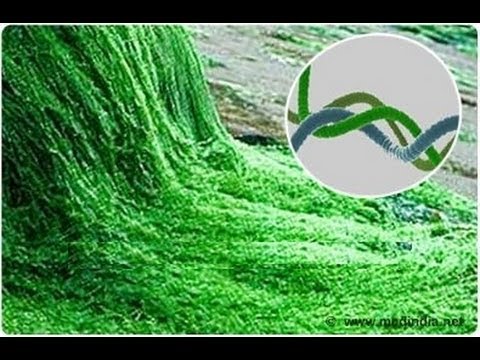
Structural Studies of Phycobiliproteins from Spirulina
Spirulina is considered to be an excellent source of proteins. The structural composition
of spirulina includes phycobiliproteins that contain a covalently attached chromophore
called phycocyanobilin, which is in a planar conformation when the protein is in native
form. When the protein is denatured, this chromophore undergoes a conformational change,
leading to a change in the absorption spectrum which is recorded at 625nm using a UV spectrophotometer.
This experiment examines the structural changes occurring in phycobiliproteins upon denaturation
with potential denaturants like urea and potassium thiocyanate, employing UV-Vis spectroscopy
Materials Required
Weighing balance Spirullina tablets
Watch glass Spatula.
100 ml beaker Sonication chamber
Ammonium Sulphate powder Magnetic stirrer
Procedure
Arrange the required materials on the Lab bench.
Preparation of Phycobiliproteins from spirullina.
Place the weighing dish over the weighing balance and tare the balance to zero.
Take four spirullina tablets open them and empty the contents into a watch glass.
Using a spatula transfer the spirullina powder from the watch glass to the watch glass kept
over the weighing balance and note the weight, which should be approximately 2g.
Transfer the powder into a 100 ml beaker with a spatula.
Using a 100ml measuring jar measure 50 ml of 0.1 Molar potassium phosphate buffer at
pH 7. Pour this into the beaker containing the spirullina
powder. Mix the solution using a glass rod.
The colour of the solution changes to deep blue colour.
The spirulina solution is then sonicated to break the cells and release the proteins.
Sonication is done 5-6 times for 1 minute at intervals of 5 minutes.
The solution is then centrifuged to remove the cell debris.
For this, the solution is transferred into the centrifuge tubes.
Centrifugation is done for 20 minutes at 24,000 RPM at 4 degree Celsius.
After 20 minutes take out the tubes from the centrifuge and you will notice blue pellets
in each tube. The supernatant is transferred into a 250
ml beaker. Take 50 ml of 0.1 Molar Potassium Phosphate
Buffer at pH 7 in a measuring jar. Make the volume of the spirullina solution
to 100 ml by adding 0.1 Molar Potassium Phosphate Buffer at pH 7 from the measuring jar.
Weigh 29.00g of ammonium sulphate powder and add it to the spirullina solution.
Mix the solution using a magnetic stirrer for 5 minutes.
After stirring, the solution is again centrifuged to isolate the proteins.
Centrifugation is done for 10 minutes at 16000 rpm at 40C.
The supernatant is transferred into a 250ml beaker and discarded, whereas the pellets
remain in each tube. Measure 25 ml buffer in a measuring jar and
add it to the pellet in each tube. Dissolve the pellet in the added buffer by
shaking the contents in the tubes with the hand.
Transfer the solution into a conical flask.
Protein Denaturation Studies.
Transfer 3mL of Potassium Phosphate buffer into a cuvette using a pipette to be used
as the blank. To a second cuvette, add 3 ml of Potassium
Phosphate buffer using a pipette. Add 100 痞 protein solutions into this cuvette
and mix it well using the pipette. Then place the cuvettes in the UV spectrometer
slots and record the spectrum at 625nm. This solution is discarded and the cuvette
is washed and dried. Now 1.5 ml potassium phosphate buffer is taken
in the cuvette. Then add the denaturant 1.50 ml of the denaturant
8M potassium thiocyanate to the cuvette. And finally add 100 痞 protein solution and
mix well using a pipette. Then place the cuvette in the spectrophotometer
slot and record the spectrum at 625nm. This solution is discarded and the cuvette
is washed well and dried. Taken 2.25ml of potassium phosphate buffer
in a cuvette . Then add 750ul of the second denaturant 8M
urea to the cuvette. Finally transfer 100 ul of protein solution
to this cuvette. Mix well using a pipette. Place the cuvette in the spectrophotometer
slot and record the spectrum at 625nm. After the measurements are completed, both
the cuvettes are removed from the UV spectrometer